Hospital Technologies
Biruni University Hospital offers advanced medical services for the diagnosis and treatment of all diseases, equipped with state-of-the-art technology in line with its vision of providing healthcare services at international standards.
Cyber Ho Magneto HoLEP Device
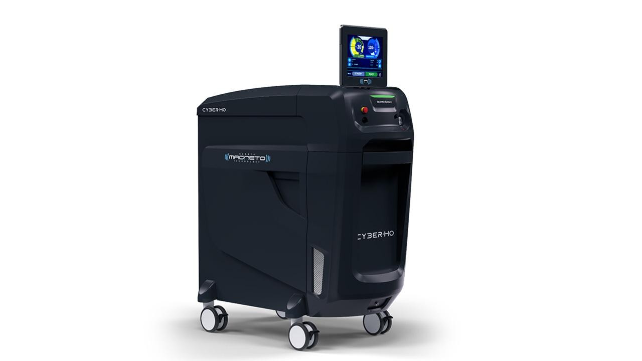
In our urology department, the Cyber Ho Magneto device, used in the treatment of benign prostatic hyperplasia (BPH), is a high-tech laser system. The system used for the treatment of prostate enlargement with HoLEP surgery offers benefits such as faster recovery and a lower risk of recurrence.
Advantages of the Cyber Ho Magneto Device
HoLEP surgery performed in our Urology department with Cyber Ho Magneto offers many benefits to our patients:
- Minimally Invasive: This method is applied by entering the body through the urinary tract without any incisions. The laser's precise tissue interaction minimizes the risk of bleeding compared to traditional surgical methods.
- Fast Recovery: The post-procedure recovery process is very fast. Our patients typically stay in the hospital for only 1-2 days and can return to their daily lives and normal activities in a short time.
- Effective Solutions for Different Sizes: Thanks to the high power and advanced technologies offered by the Cyber Ho Magneto, HoLEP surgery can be safely and extremely effectively applied to small, medium, or very large prostates.
- Low Risk of Recurrence: With the HoLEP method, the enlarged tissue (adenoma) that obstructs urine flow is completely removed. Therefore, the likelihood of the disease recurring in the future is extremely low.
- Preservation of Sexual Functions: The laser's precise and controlled effect on tissues minimizes the risk of damaging the nerves related to sexual functions around the prostate.
Cios Select with FD
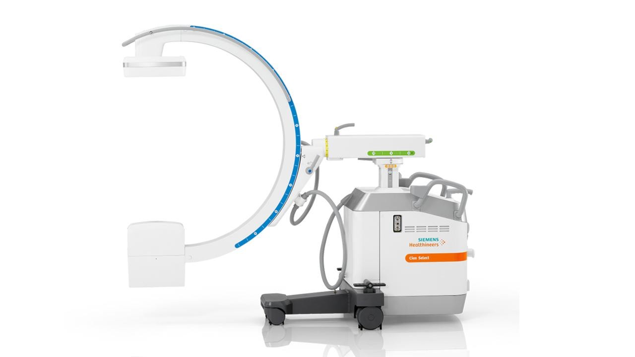
The Cios Select with FD is a mobile C-arm device featuring flat-panel detector (FPD) technology. This technology provides high-resolution and clear images, ensuring greater precision in surgical procedures.
What are the Advantages of the Cios Select with FD?
- Superior Image Quality: Flat-panel detector technology provides clearer and more detailed images than traditional devices. This allows surgeons to see anatomical structures much better and perform more precise interventions.
- Low Radiation Dose: Cios Select with FD offers the possibility of working with a lower radiation dose for patients and healthcare personnel, thanks to its advanced dose optimization features.
- Wide Range of Applications: Suitable for use in many different fields, such as orthopedics, traumatology, general surgery, and urology.
- User-Friendly Interface: Its smart touchscreen and intuitive user interface allow for easy and fast operation.
- Mobile Design: Its compact and lightweight structure allows it to be easily transported and used in different operating rooms.
MAMMOMAT Revelation
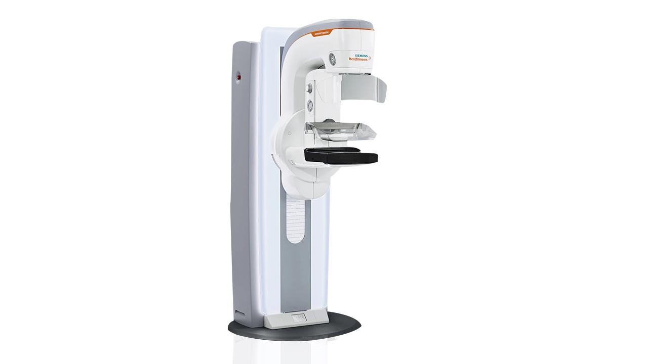
At Biruni University Hospital, we stand with you against breast cancer with our MAMMOMAT Revelation device. This mammography device, with its 50-degree wide-angle tomosynthesis technology, obtains 3D images of breast tissue, ensuring clearer and more accurate detection of lesions. This makes it possible to diagnose and begin treatment for breast cancer in its early stages.
Advantages of MAMMOMAT Revelation
The MAMMOMAT Revelation 3D mammography device offers the following advantages in breast cancer screening:
- Higher Accuracy: Wide-angle tomosynthesis technology provides higher accuracy in cancer detection.
- Earlier Diagnosis: It allows for the detection of even small lesions in their early stages, increasing treatment success.
- More Accurate Results: The clear separation of overlapping tissues reduces the number of false-positive results and minimizes the need for unnecessary biopsies.
- Less Radiation: Thanks to PRIME technology, up to 30% less radiation dose is used compared to standard 2D mammography.
- A More Comfortable Experience: Personalized soft compression technology reduces the discomfort patients feel during compression. AEC (Automatic Exposure Control) and OpDose features make it possible to use the most suitable amount of radiation for each woman.
The Mammomat Revelation mammography device enables the early diagnosis of breast cancer that has not yet shown symptoms. It is also used for detailed examination and diagnosis if a suspicious mass or finding is detected in the breast.
The device combines images taken from different angles of the breast tissue to create a 3D image, which helps to reach the suspicious area more accurately during a biopsy. It also helps to visualize lesions more clearly.
MAGNETOM Vida 3 Tesla MR
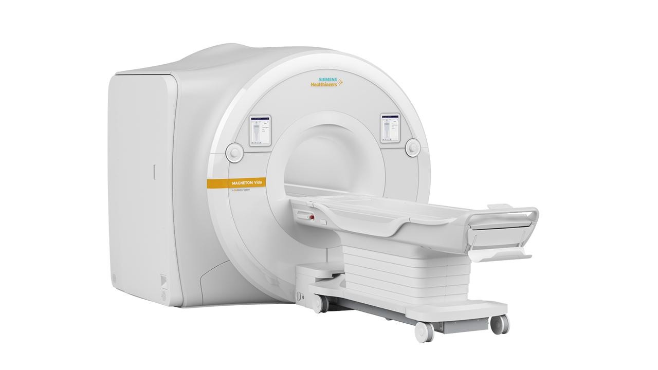
Magnetic Resonance Imaging (MRI) is a non-invasive method that uses magnetic fields and radio waves to create detailed images of the body's internal structures. The MAGNETOM Vida BioMatrix 3 Tesla MRI device in our hospital is twice as powerful as standard 1.5 Tesla MRI devices in terms of magnetic field strength. This high magnetic field allows for the acquisition of more detailed, high-resolution images in thinner slices.
Advantages of the MAGNETOM Vida BioMatrix 3 Tesla MRI Device
MAGNETOM Vida BioMatrix is an MRI device that prioritizes patient comfort while also offering superior image quality. Here are the prominent features of this device:
- BioMatrix Technology: It includes smart sensors that adapt to the patient's body shape. These sensors make automatic adjustments based on the patient's body measurements and shape, providing personalized imaging. It offers optimized imaging, especially for patients with obesity or different body types.
- Clearer and More Accurate Images: With a 3 Tesla magnetic field strength, it enables detailed imaging of complex structures such as the brain, spine, joints, and soft tissues. It offers superior diagnostic accuracy in detecting small lesions and early-stage diseases.
- High Patient Comfort: With a wide patient entry and short tunnel structure, it minimizes the feeling of claustrophobia. The soft-surfaced patient bed and quiet operation feature ensure a comfortable experience for patients.
- Fast and Reliable Results: Thanks to advanced software and hardware features, scan times are shortened while maintaining image quality. It provides the opportunity for quick diagnosis in emergency situations.
- Versatile Use: It can be used in many areas, such as neurological, orthopedic, cardiac, oncological, and abdominal imaging. It supports advanced imaging techniques like diffusion, perfusion, spectroscopy, and functional MRI.
In What Cases Is 3 Tesla MRI Used?
The MAGNETOM Vida BioMatrix 3 Tesla MRI device can be used in many different fields, from the detection of cancer tumors to the detailed examination of heart valves:
- Neurological Imaging: Diagnosis and follow-up of neurological diseases such as brain tumors, stroke, multiple sclerosis, Alzheimer's, and Parkinson's.
- Orthopedic Imaging: Evaluation of joint injuries, meniscus tears, spine diseases, and cartilage damage.
- Cardiac Imaging: Detailed examination of the heart muscle, valves, and vascular structures.
- Oncological Imaging: Detection, staging, and follow-up of cancer tumors.
- Abdominal Imaging: Detailed imaging of the liver, kidneys, pancreas, and other internal organs.
Biruni University Hospital, located in Küçükçekmece, Istanbul, is at your service with state-of-the-art medical devices. Our 3 Tesla MRI device, with its patient-focused design and superior technology, offers a unique experience for both patients and doctors. It ensures faster, more comfortable, and more accurate imaging processes.
SOMATOM Force CT Device
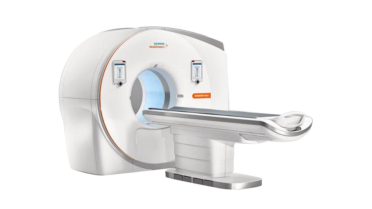
The SOMATOM Force CT device is at your service at Biruni University Hospital, revolutionizing the field of computed tomography with high speed, low radiation dose, and superior image quality.
SOMATOM Force CT is the world's first dual-source and high-energy computed tomography device. The device offers much faster scan times, a lower radiation dose, and unparalleled image clarity compared to traditional CT devices. It provides unique performance, especially in imaging moving organs (heart, lungs) and complex anatomical structures.
Advantages of the SOMATOM Force CT Device
The prominent features of the SOMATOM Force computed tomography device are as follows:
- Shorter Scan Time: Two X-ray tubes and two detector systems work simultaneously, reducing the scan time by 10 times. It provides high temporal resolution for imaging moving organs like the heart. In addition, a full-body scan can be performed in the time of a single breath-hold.
- Low Radiation and Contrast Agent: The radiation dose is reduced by up to 60% with ADMIRE (Advanced Modeled Iterative Reconstruction) technology. Unnecessary radiation absorption is prevented with the Tin Filter. In addition, the use of a contrast agent is reduced by up to 50%, which lightens the load on the kidneys.
- Superior Image Quality: It is possible to detect even micro-lesions with thin slices of up to 0.4 mm. The wide gantry opening of 78 cm provides a comfortable imaging experience for all patients, including those with obesity. With the Spectral Imaging (Dual-Energy) feature, the chemical components of tissues can be analyzed. This allows structures such as tumors, gout, or vascular plaques to be clearly distinguished.
- Patient-Friendly Design: Patient stress is minimized with silent operation technology. The soft and wide patient table provides comfort, especially during long scans.
In Which Areas Is the SOMATOM Force Computed Tomography Used?
- Cardiological Imaging: Non-invasive evaluation of coronary artery diseases, heart valves, and bypass grafts. Obtaining clear results in patients with arrhythmia by imaging independently of heart rate.
- Oncological Evaluation: Follow-up of tumor size, metastases, and treatment response. Analysis of the metabolic activity of the tumor.
- Emergency and Trauma: Fast diagnosis in emergency cases (brain hemorrhage, internal organ injuries) thanks to very fast scan times.
- Pediatric Imaging: Safe use in pediatric patients with a low radiation dose.
- Neurological and Vascular Examinations: Detailed analysis of cerebral, carotid, and peripheral vessels.
The SOMATOM Force CT device is an advanced computed tomography device that enables the high-accuracy detection of micro-lesions and early-stage diseases. It offers a life-saving solution in emergency interventions with scans completed in seconds, while protecting your organs with radiation and contrast agent dose optimization.
Mammomat Inspiration Mammography Device
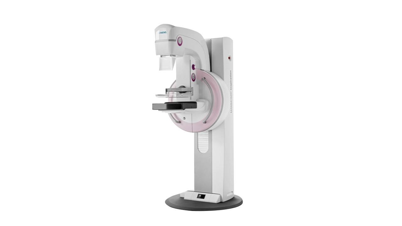
In our hospital, we use the Mammomat Inspiration digital mammography device for the early diagnosis of breast cancer. This advanced technology device makes the screening process safe and effective by combining high image quality, low radiation dose, and patient comfort.
Advantages of the Mammomat Inspiration Mammography Device
The advantages of the MAMMOMAT Revelation 3D mammography device in breast cancer screening are as follows:
- Higher Accuracy: Wide-angle tomosynthesis technology provides higher accuracy in cancer detection.
- Earlier Diagnosis: It allows for the detection of even small lesions in their early stages, increasing treatment success.
- More Accurate Results: The clear separation of overlapping tissues reduces the number of false-positive results and minimizes the need for unnecessary biopsies.
- Less Radiation: Thanks to PRIME technology, up to 30% less radiation dose is used compared to standard 2D mammography.
- A More Comfortable Experience: Personalized soft compression technology reduces the discomfort patients feel during compression. AEC (Automatic Exposure Control) and OpDose features make it possible to use the most suitable amount of radiation for each woman.
The Mammomat Revelation mammography device enables the early diagnosis of breast cancer that has not yet shown symptoms. It is also used for detailed examination and diagnosis if a suspicious mass or finding is detected in the breast.
The device combines images taken from different angles of the breast tissue to create a 3D image, which helps to reach the suspicious area more accurately during a biopsy. It also helps to visualize lesions more clearly.
MOBILETT Elara Max X-Ray Device
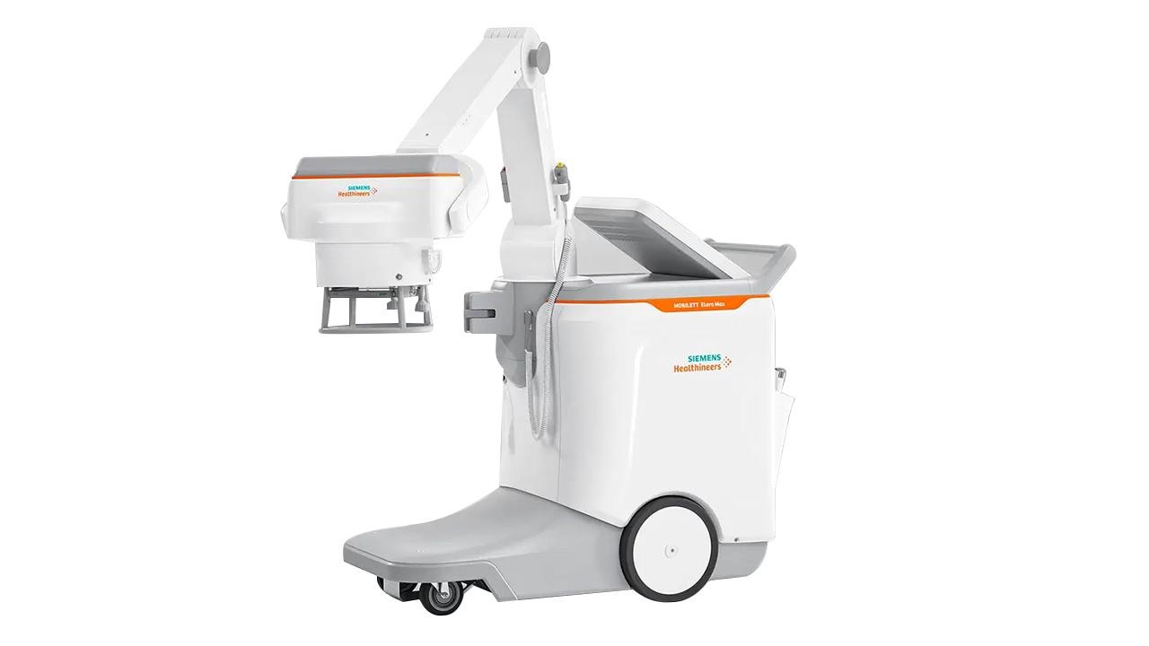
The MOBILETT Elara Max is a mobile X-ray device that stands out with its superior maneuverability, high-resolution imaging, and fast results. It provides fast and reliable diagnostic capabilities, especially in dynamic environments such as intensive care units, operating rooms, and emergency departments.
Advantages of the MOBILETT Elara Max X-Ray Device
- Superior Maneuverability: Thanks to its compact design and advanced wheel system, it can be easily moved even in narrow corridors and crowded patient rooms. This provides great convenience in meeting the X-ray imaging needs of bedridden patients.
- High-Resolution Imaging: It offers excellent image quality with its advanced detector technology and automatic exposure control. Sharp and detailed images support clinical decision-making processes and help to achieve an accurate diagnosis.
- Fast Diagnosis: It provides the opportunity for fast imaging in emergencies, thus allowing for timely intervention and treatment.
- Radiation Dose Reduction: It minimizes patients' exposure to radiation through optimum dose control.
- Comfortable Patient Care: It offers bedside imaging services for bedridden individuals, preventing unnecessary patient transfers and discomfort.
- Infection Control: Easily cleanable surfaces and an antibacterial coating minimize the risk of infection and increase your safety.
As Biruni University Hospital, we have added the MOBILETT Elara Max X-ray device to our mobile X-ray fleet as part of our commitment to providing excellence in patient care and adopting the latest developments in medical technology. For detailed information and appointments, you can call 444 8 276.
Artis Icono Biplane Angiography Device
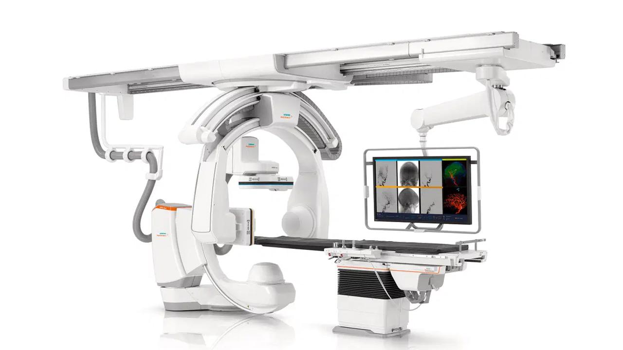
The Artis Icono Biplane angiography device is a highly advanced imaging angiography system designed for hybrid operating rooms and minimally invasive procedures. With high precision, flexibility, and a user-friendly interface, it offers real-time and detailed imaging.
Advantages of the Artis Icono Biplane Angiography Device
- Better Images: The device's OPTIQ imaging technology makes it possible to obtain high-resolution and clear images with a low dose of radiation. In addition, its 3D imaging feature allows for the detailed visualization of anatomical structures.
- Faster Recovery: Artis Icono Biplane allows for less invasive procedures. This means you can have your treatment with smaller incisions and recover faster. This way, you can spend less time in the hospital and quickly return to your daily life.
- Less Pain: Minimally invasive procedures cause less pain and discomfort. Post-operative pain is usually mild, and your recovery process is comfortable.
- Lower Risk: With advanced imaging technology, your doctors can perform procedures more precisely. This reduces the risk of complications and increases the chance of treatment success.
- Less Radiation: Artis Icono Biplane provides imaging using a low dose of radiation. This minimizes your risk of radiation exposure.
- More Comfortable: The system offers a more comfortable treatment experience with its ergonomic design and silent operation feature.
Artis Icono Biplane Application Areas
- Cardiovascular Procedures: Procedures such as coronary angiography, balloon angioplasty, stent placement, and valve repair and replacement.
- Neurovascular Procedures: Procedures such as the treatment of brain aneurysms, AVM embolization, and acute ischemic stroke treatment.
- Peripheral Vascular Procedures: Procedures such as the treatment of narrowing or blockages in the arteries of the legs and arms.
- Oncological Procedures: Procedures such as tumor embolization, chemotherapy applications, and radiofrequency ablation.
Artis Zee Pure Angiography Device
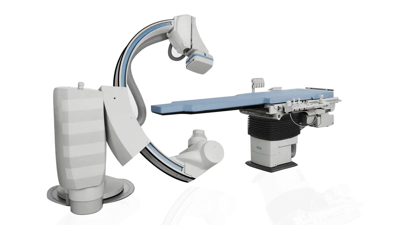
Artis Zee Pure is a versatile angiography system designed especially for cardiac and vascular procedures. Thanks to its advanced imaging technologies and innovative features, it offers a safer, faster, and more comfortable treatment experience.
Advantages of the Artis Zee Pure Angiography Device
- High Image Quality: Artis Zee Pure enables the acquisition of detailed and clear images of the cardiovascular system thanks to its high-resolution imaging technology and advanced image processing algorithms. This allows your physician to determine the cause of your heart condition more accurately and plan the most suitable treatment for you.
- Minimal Radiation Dose: Artis Zee Pure is equipped with advanced dose management tools such as CARE and PURE. These tools help minimize your radiation exposure.
- Speed and Flexibility: The robotic C-arm of the Artis Zee Pure angiography system allows for faster positioning and a wider anatomical reach. This means your procedures are completed in a shorter time, and you spend less time in the hospital
- Advanced Clinical Applications: Artis Zee Pure supports advanced clinical applications such as stent optimization, fractional flow reserve (FFR) measurement, and 3D imaging. These applications allow for more precise diagnosis and treatment.
MultiX Impact X-ray Device
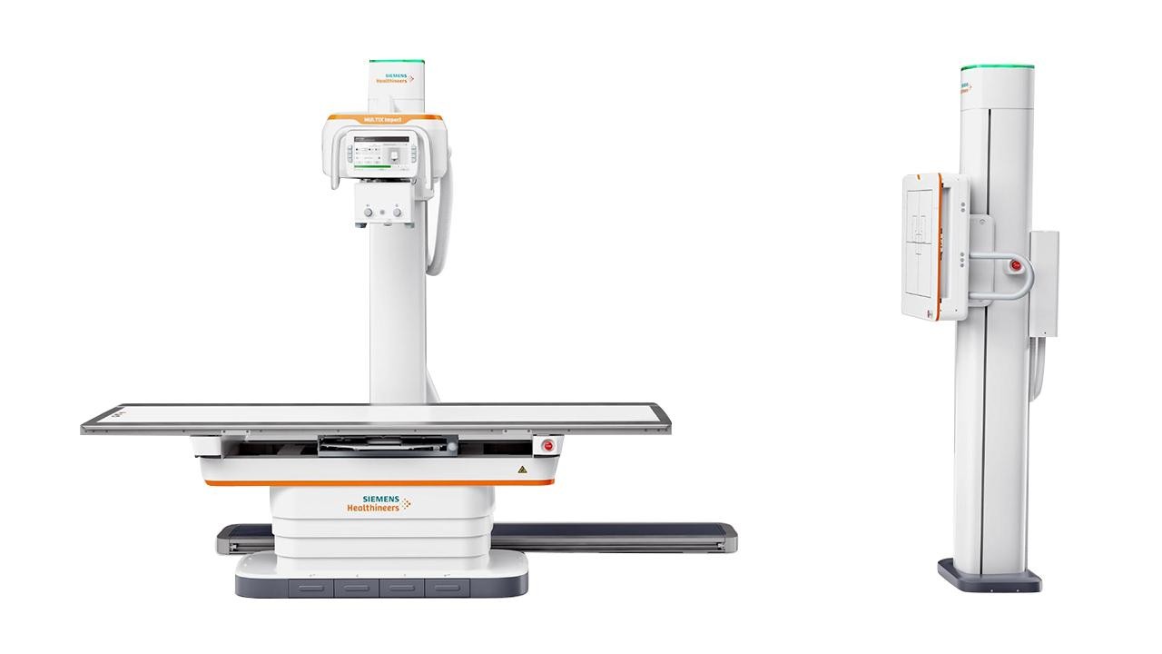
The MultiX Impact X-ray device is designed to meet modern medical imaging needs. The device combines high-quality imaging, a low radiation dose, and patient comfort.
Advantages of the MultiX Impact X-ray Device
- Low Radiation Dose: MultiX Impact is much safer than traditional X-rays with its low radiation dose. This technology minimizes radiation exposure, especially for children, pregnant women, or patients who need frequent X-rays.
- Results in 10 Seconds: Images are instantly displayed on the digital screen. This reduces the need for re-shooting, and your treatment plan can begin quickly.
- Artificial Intelligence Support: The device automatically optimizes images to provide clear results. We aim for the best quality for your doctor to make an accurate diagnosis.
- Comfortable Positioning: With the height-adjustable patient table, you can have your X-ray comfortably while sitting, lying down, or even in a wheelchair.
- Wireless Detector: It provides you with freedom of movement even in painful areas and difficult positions.
SOMATOM Go.Now CT Device
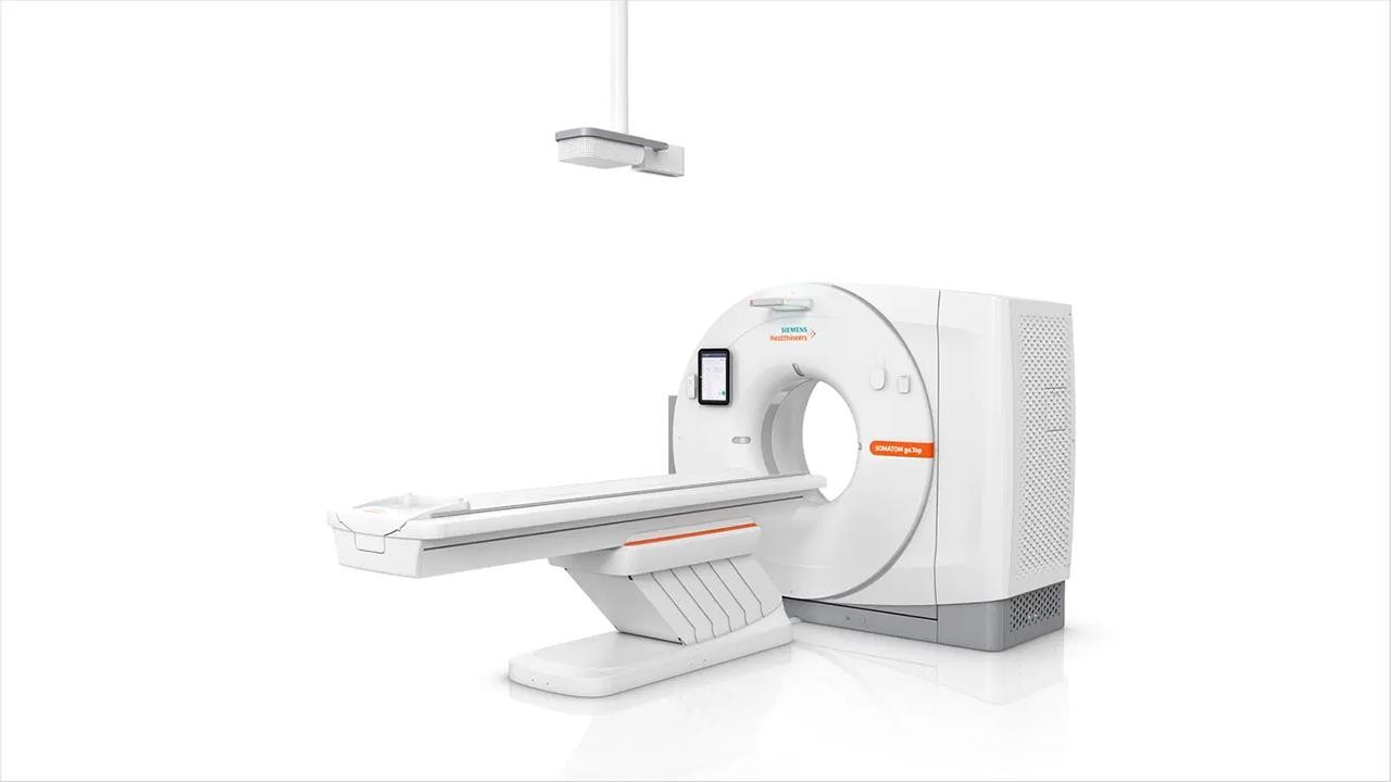
SOMATOM Go.Now is a next-generation computed tomography (CT) device. It allows for the acquisition of fast and high-quality images with low radiation. The device, which stands out with its design that provides high patient comfort, has a wide range of uses from cardiology to gastroenterology.
Advantages of the SOMATOM Go.Now CT Device
- Fast and Efficient: Its fast scanning feature allows for the acquisition of high-quality images in a short time. The FAST Integrated Workflow feature optimizes the scanning process and reduces the time you need to spend in the device.
- Low Radiation Dose: Our patients' safety is our priority. By working with a low radiation dose, we minimize your risk of radiation exposure. This feature is of great importance, especially for children and individuals who require repeated scans.
- Superior Image Quality: Thanks to its advanced technology, it is possible to obtain high-resolution and detailed images. This helps our physicians make a more precise diagnosis.
- Wide Range of Applications: The device can be used to image different parts of the body (head, neck, chest, abdomen, spine, etc.). It is also suitable for advanced applications such as angiography.
- Patient Comfort: The design of the device has been made with your comfort in mind. The wide and bright scanning area helps you have a comfortable tomography experience.
In What Cases Is the SOMATOM Go.Now CT Device Used?
The SOMATOM Go.Now CT device can be used in the diagnosis of various medical conditions:
- Oncology: Cancer diagnosis, tumor staging, treatment planning, and follow-up.
- Cardiology: Diagnosis of heart diseases such as coronary artery disease, heart valve diseases, and heart failure.
- Neurology: Diagnosis of neurological diseases such as brain tumors, stroke, and MS.
- Orthopedics: Diagnosis of musculoskeletal diseases such as fractures, dislocations, joint diseases, and spine problems.
- Gastroenterology: Diagnosis of digestive system diseases.
- Urology: Diagnosis of kidney and urinary tract diseases such as kidney stones and urinary tract infections.
- Emergency Medicine: For quick diagnosis in emergency situations such as traffic accidents and falls.
PET-CT Device
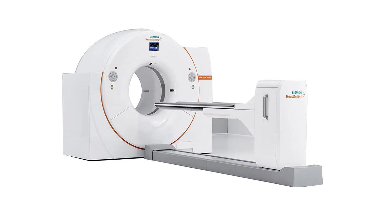
PET-CT, also known as Positron Emission Tomography (PET) and Computed Tomography (CT), is a hybrid imaging technology that combines both imaging methods. It shows metabolic activity and anatomical structure simultaneously, allowing for earlier and more accurate diagnosis of diseases.
Advantages of the PET-CT Method
PET-CT scanning is the gold standard method used in the diagnosis and treatment of many diseases, such as cancer, heart disease, neurological disorders, and infections. Its advantages include:
- Early Diagnosis: It makes it possible to diagnose diseases in their earlier stages. This is very important for the success of the treatment.
- Precise Diagnosis: It allows for a more precise diagnosis compared to other imaging methods.
- Treatment Planning: It allows for more effective treatment planning.
- Treatment Monitoring: It helps in evaluating the response to treatment and monitoring the disease.
Our hospital uses the General Electric Discovery IQ device. The device offers fast scanning with a low dose, thanks to its NEMA sensitivity, motion correction feature, and four-ring cycle.
SPECT-CT Device

SPECT-CT, also known as Single Photon Emission Computed Tomography (SPECT) and Computed Tomography (CT), is a hybrid system that combines both imaging methods.
SPECT measures the functions and metabolic activities of the organs in your body. By injecting a special drug, it provides a detailed image of how the target organ or tissues are working. CT, on the other hand, provides detailed images of your body's anatomical structure.
Advantages of SPECT-CT
By combining two powerful imaging methods, SPECT-CT helps in the diagnosis and treatment of many diseases, covering many fields from cardiology to neurology, endocrinology to oncology. The advantages are as follows:
- Accurate and Reliable Diagnosis: It gets to the root of your disease and shows you and your physician the most accurate treatment path.
- Early Diagnosis: It increases your chances of treatment by detecting your disease in its earlier stages.
- Personalized Treatment: It allows for the creation of a personalized treatment plan for you.
- Treatment Process Monitoring: It guides you on your recovery journey by monitoring the effectiveness of your treatment.
- Low Radiation Dose: It prioritizes your safety with a lower radiation dose compared to traditional methods.
- Fast and Comfortable Scan: It offers a comfortable experience with short-duration scans and a relaxed environment.
Gamma Probe (Handheld Gamma Detector)
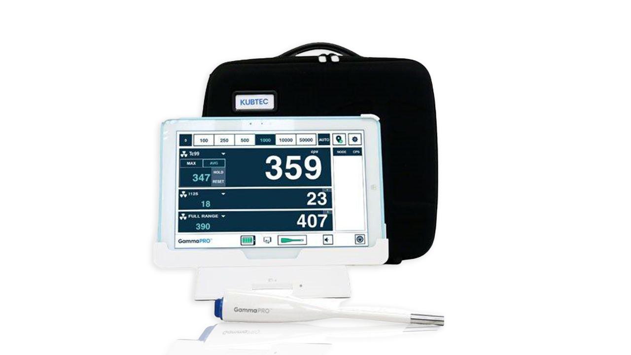
The Gamma Probe is a portable nuclear medicine detector used, especially during surgery or at the bedside. It is used to detect radioactively labeled tissues or lymph nodes. It helps to locate the target tissue directly during surgery.
Main Application Areas
- Sentinel Lymph Node Biopsy: Locating the first lymph node to which a tumor may have spread in breast cancer or melanoma surgery.
- Thyroid Surgery: Determining the location of thyroid tissue or nodules.
- Radioactive Tumor Localization: Finding radioactively labeled areas in rare tumors.
- Parathyroid Surgery: Detecting parathyroid glands during surgery.
Advantages
- Real-time radioactivity detection.
- Shortening surgery time.
- Increasing the success rate of surgery.
- Enabling minimally invasive procedures.